Breaking Down Excellence: How Can Top Medical Imaging Services Improve Diagnostic Accuracy?
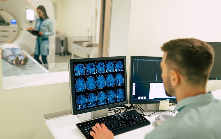
Because it enables physicians to view the inside organs and systems of the patient without intrusive procedures, medical imaging is essential to the delivery of contemporary healthcare. Modern medical imaging methods including PET, CT, MRI, and ultrasound scans offer comprehensive functional and anatomical data that helps with diagnosis and directs therapy choices. New difficulties are brought about by the vast amount and complexity of medical images that are produced every day. To solve this, top medical imaging services are improving diagnostic accuracy by utilizing cutting-edge technology and knowledgeable analysis.
Cutting Edge Technology for Exceptional Image Quality
The newest imaging gear and software are used by top medical imaging services to produce high-resolution images with superb contrast and clarity. Modern CT and MRI scanners can visualize previously impossible-to-see anatomical features. For a thorough understanding of pathology, radiologists can manipulate multi-planar reconstructions and three-dimensional renderings with powerful workstations that are outfitted with specialist visualization tools.
Additionally enhancing image quality are machine learning technologies and artificial intelligence. Deep neural network-powered denoising and artifact reduction approaches result in clearer scans with less noise and artifacts. Sophisticated reconstruction techniques reduce radiation exposure while optimizing spatial and contrast resolution. Consistency across locations and equipment is ensured by automated quality control. When taken as a whole, these technical developments give radiologists better-quality images for precise diagnosis.
Sub-Specialization in Focused Subject Matter
Prominent medical imaging companies understand that every case is unique. They create sub-specialized teams with a focus on particular therapeutic areas, such as cardiology, musculoskeletal imaging, oncology, and more, to improve diagnostic precision. Sub-specialist radiologists are trained in advanced imaging modalities, diseases, and protocols specific to their field of specialization. They have a deeper comprehension of how diseases present, how treatments work, and how subtle imaging results are.
Subspecialists can identify small anomalies earlier and provide a more thorough description of them thanks to their focused knowledge. To make the best possible management decisions, they can link imaging results with clinical data and uncover important imaging biomarkers. Effective diagnosis and treatment planning are facilitated by multidisciplinary teams that comprise radiologists, oncologists, surgeons, and other professionals. Subspecialization makes use of medical imaging’s precision in describing and tracking disease.
Consistent Procedures and Quality Control
Quality assurance procedures and uniform, defined processes are characteristics of top-tier medical imaging services. Scan settings are optimized, pertinent anatomy is covered, and comparative studies are produced over time with well-defined protocols. Consistency and productivity are further improved by automatically selecting protocols based on clinical indications.
Peer review of scans, phantom imaging for accuracy and precision assessment, routine equipment calibration, and key performance metrics monitoring are all components of robust quality control. Protocols are strengthened when incidents are learned from inconsistencies or overlooked findings. The retrieval of pertinent previous studies and thorough documentation are made easier by the use of organized data capture and reporting templates.
Standardized procedures and high-quality control methods reduce variation and optimize diagnostic efficacy between locations and technicians. They set benchmarks to monitor gains in performance and guarantee a high and constant level of care.
Superior Visualization and Guidance Support Instruments
Modern visualization techniques are revolutionizing radiologists’ analysis of medical pictures. Multi-planar viewing, three-dimensional volume rendering, dynamic contrast enhancement curves, and the merging of complementary modalities like PET/CT are all made possible by advanced workstations.
Applications of artificial intelligence are improving visualization. Anatomical structures are delineated by automated segmentation technologies to facilitate interpretation. Quantitative imaging biomarkers associated with diagnosis and prognosis are using image processing techniques such as radionics, deep learning, and texture analysis.
Applications for decision support combine clinical information and imaging results to produce differential diagnoses, calculate prognosis ratings, and suggest evidence-based treatment alternatives. Some programs even help radiologists by automatically detecting lesions.
By utilizing medical pictures and related data, these cutting-edge visualization and decision-support tools can improve clinical relevance and diagnostic confidence. They enable radiologists to interpret scans more and tailor recommendations for patient care.
Professional Agreement for Coherent Interpretation
Even though technology facilitates analysis, a skilled radiologist is still essential to the diagnosis process. Prominent medical imaging systems encourage continual education and multidisciplinary consensus discussions to foster consistent, competent interpretation.
Using weekly tumor boards and morbidity and mortality conferences, subspecialists can collaborate to review difficult cases. This makes it easier for peer review to identify differences and creates agreement on the best course of action. It also acts as a forum for talking about the most recent research updates and correlations between imaging and pathology.
Radiologists are guaranteed to retain their expertise through continuing medical education that covers the most recent methods, procedures, and clinical applications. Proctoring programs and fellowships aid in bringing uniformity to the team’s knowledge and skill set.
Combining these consensus-building and educational initiatives assists radiologists in reaching consistent, fact-based conclusions. To reduce reporting variability, they create an understanding of normal variants and disease manifestations. maximizing diagnostic accuracy, expert consensus interpretation must be of the collective clinical experience.
The field of medical imaging has a lot of potential to improve diagnosis accuracy in the future. Human anatomy and physiology will be shown in even greater depth thanks to emerging technologies like molecular imaging, 3D bioprinting, augmented and virtual reality, and artificial intelligence. Innovative uses of computer vision, machine learning, and natural language processing will facilitate the automation of repetitive processes and the discovery of new imaging biomarkers. By linking genes and imaging characteristics, radio genomics will aid in the prediction of treatment outcomes. Large-scale research collaborations will be power by sharing de-identified image data made possible by blockchain and cloud-based technology. These fascinating developments will continue to push medical imaging to new heights and move us closer to universally available, individualized, precision-based treatment if they are responsible and for the good of patients.
Conclusion
Leading best medical imaging analysis are improving diagnostic precision in novel ways by fusing state-of-the-art technology with focused expertise, standardized procedures, sophisticated visualization tools, and expert consensus. By accurately diagnosing and tracking diseases, they optimize and personalize care by utilizing the full potential of medical images. With the ongoing development of artificial intelligence and imaging technologies, these services will find new ways to provide precision healthcare and gain deeper clinical insights.

